Microscopic Imaging Spectroscopy System
MISM-05 |
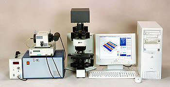 |
This system attaches to an optical microscope to perform microspectroscopy and imaging spectroscopy. |
■Features |
- Enables microspectroscopic measurements of spots as small as 1 μm
- Supports spectral, transmittance, absorbance, and reflectance measurements in microscopic imaging spectroscopy
- Enables imaging spectroscopy with a wavelength resolution of 1 nm
|
■Measurement Principle |
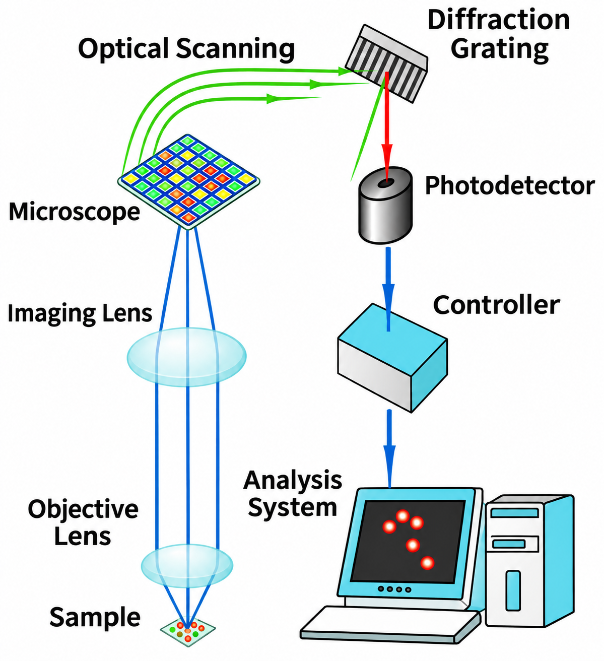 |
A magnified image of the sample is formed by an objective lens and an imaging lens. This image is optically divided into approximately 50 μm pixels and scanned. Light from each pixel passes through a spectrometer (diffraction grating), which selects the desired wavelength for measurement by a photodetector. A dedicated controller controls the optical scan and processes the photodetector signals. The resulting spectral image is then viewed using the analysis system. Microspectroscopic measurements can also be performed at any selected point within the spectral image. |
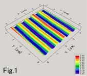 |
The 3D image in Fig. 1 shows a Ronchi ruling resolution test target for microscopy. Its spatial frequency is 600 lines/mm, and its line width is approximately 0.8 μm. These data indicate that the system provides high image resolution.
The analysis system provides the following functions for both microspectroscopy and imaging spectroscopy:
1. Spectral measurement
2. Transmittance measurement
3. Absorbance measurement
4. Reflectance measurement |
|
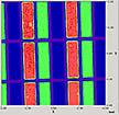
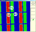
Fig. 2 shows a monochrome image of a liquid crystal display color filter at 480 nm.
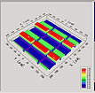
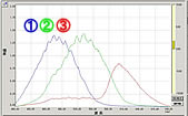
Fig. 3 shows a 3D image with light intensity plotted along the Z-axis. As shown in Fig. 4, any point in the monochrome image can be selected for spectral analysis of that microscopic location (see Fig. 5). |
 |
|
 |
|
 |
|
 |
| Fig. 2 |
|
Fig. 3 |
|
Fig. 4 |
|
Fig. 5 |
|
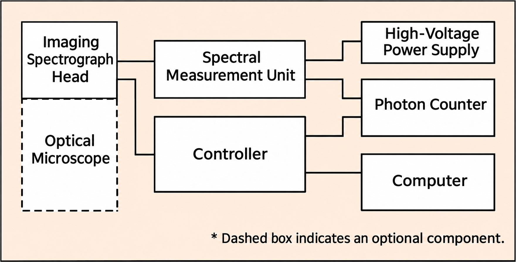
■System Configuration |
 |
■Specifications |
| Measurement Wavelength Range |
350 nm–700 nm |
| Wavelength Resolution |
1 nm *1 |
| Image Resolution |
1 μm *2 |
| Light-Receiving Area |
15 mm × 15 mm |
| Detector Element Size |
10 μm or 50 μm |
| Spectrometer Focal Length |
100 mm |
| Photodetector |
Photomultiplier tube |
| Photon Counting Pulse Resolution |
10 ns |
| Gate Time |
50 μs–60 s |
|
*1 Wavelength resolution depends on the measured light intensity.
*2 Image resolution depends on the resolution of the connected microscope. |
*Specifications and appearance are subject to change without notice for product improvement. |
| |

